

Left Ventricular Modelling
Mathematical modelling of left ventricular still remains a great challenging due to its multi-scale, multi-physics and subject-specific nature.
- Multi-scale: cellular levle calcium dynamics, myocyte active contraction, ...
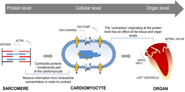
Figure 1, Illustration of multi-scale nature of heart mechanics
- Multi-physics: blood flow mechanics (fluid mechanics), nonliear soft tissue mechanics (solid mechanics), electrophysiological activities, etc, and their interactions, ...
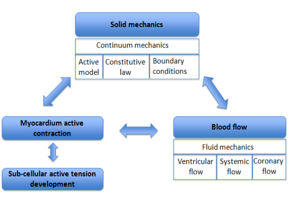
Figure 2, Illustration of multi-physics nature of heart mechanics
- Subject-specific: goemetry, boundary conditions, material parameters (active and passive), ...
We are using a recent version of immersed boundary method developed by Boyce E. Griffith from New York Univeristy, USA for subject specific left ventricle modelling, from clinical images to in-sillico models, from cellular level active contraction to LV pumping, etc. The followed animation shows the LV model is contracting from end of diastole to end of systole for a healthy subject, superimposed with LVOT CMR images, the model is working well compared the in-vivo images.
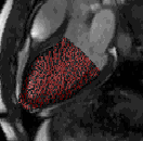
Figure 3, deformed LV endocardial surface from end-diastole to end-systole
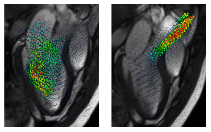
Figure 4, a numerical simulation of a healthy young male heart. Left: vortex forming inside ventricular chamber during diastole; right: strong jet flow in the aorta during systole.